How some patients came to me, what we found, what treatment involved, and how they did afterwards. Shared to help other patients understand their condition.
Each case is shared with the patient’s written consent, with names and identifying details removed. Every eye is different: these cases explain what treatment can involve and do not promise the same result for anyone else.
Reading the vision numbers: 6/6 is normal distance vision; the bigger the second number, the weaker the vision (6/60 is very poor). N6 is normal reading-size print. Tap any scan to see it larger.
Retina surgeryMacular hole surgery
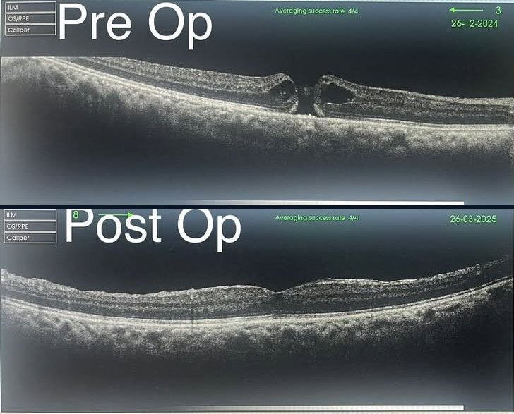
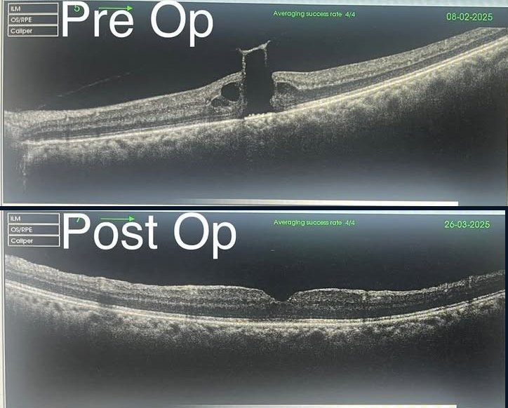
Macular holes in both eyes, repaired one month apart
60s · Male · 2024–2025
PresentationBlurred and distorted central vision in both eyes. Straight lines looked bent, and faces and small print were hard to see.
HistoryAged 63. Both eyes were affected.
FindingsOCT scans showed a full-thickness macular hole in each eye, a small break in the centre of the retina. Vision was 6/36 in the right eye and 6/60 in the left.
TreatmentVitrectomy surgery to close the hole, first in the right eye and one month later in the left.
OutcomeWithin a month of each operation the holes had closed on OCT. Vision improved from 6/36 to 6/9 in the right eye and from 6/60 to 6/12 in the left.
Right eye: before surgery (top) and after (bottom)Left eye: before surgery (top) and after (bottom)
What patients can learn from thisA macular hole can often be closed with surgery, and vision can improve a great deal, even when both eyes are affected. If your central vision becomes blurred or distorted, have your retina checked early.
Retina surgeryVitrectomy with membrane peel
Blurred vision after a successful cataract operation: an epiretinal membrane
60s · Female · 2024–2025
PresentationVision in the right eye slowly became blurred a few months after cataract surgery.
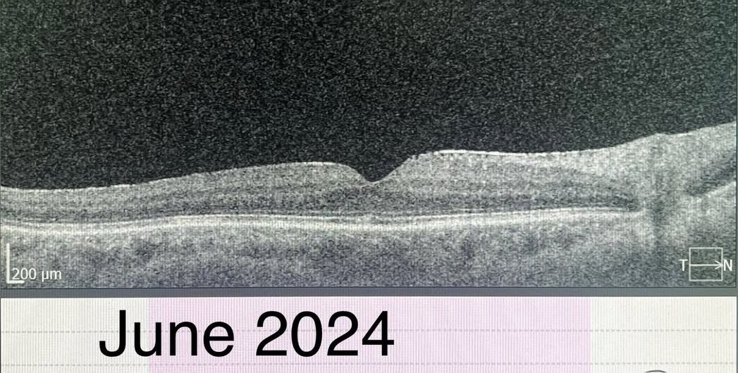
HistoryAged 68. Cataract surgery on the right eye in June 2024, after which vision was perfect (6/6 distance, N6 near).
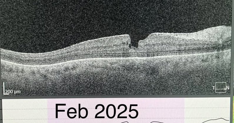
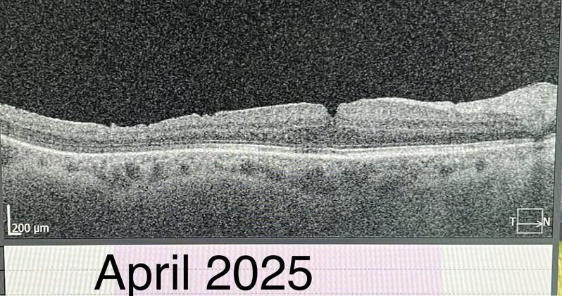
FindingsIn February 2025 vision had dropped to 6/12 and N8. An OCT scan showed an epiretinal membrane: a thin layer of scar tissue on the surface of the retina, wrinkling it and blurring vision.
TreatmentPars plana vitrectomy with peeling of the membrane, February 2025.
OutcomeBy April 2025 vision had returned to 6/6 and N6.
June 2024: normal retina after cataract surgeryFebruary 2025: epiretinal membrane before surgeryApril 2025: after membrane peel
What patients can learn from thisIf vision gets worse months after a good cataract operation, the cause may be in the retina rather than the new lens. An OCT scan can find it, and surgery can restore vision.
Have similar symptoms?
Book a consultation so your own eyes can be examined. Treatment depends on your examination and tests.